HIP DYSPLASIA

CANINE HIP DYSPLASIA
-disease of coxafemoral joint which lead to degeneration of articular cartilage , dvlp of osteoarthritis
 abnormal formation of the hip socket that, in its more severe form, can eventually cause crippling lameness and painful arthritis of the joints. 

During growth, both the ball (the head of the femur, or thighbone) and the socket in the pelvis (acetabulum) must grow at equal rates. In hip dysplasia, this uniform growth during puppyhood does not occur. The result is laxity of the joint, followed by degenerative joint disease (DJD) or osteoarthritis (OA), which is the body's attempt to stabilize the loose hip joint.

TARGET-large breed 
 first affected group is the adolescent dogs, typically six to 18 months of age.The radiograph on the right above shows the hips of such a patient. This dog has hip dysplasia but has not yet developed arthritis.

Many dogs with similar radiographs will not be in pain and thus will not end up coming in for an evaluation. These dogs show up later as elderly dogs, after they have been walking on their poorly formed hips for many years. After many years, bony spurs along the margins of the socket, mineralization of the joint capsule, cartilage wear, and inflammatory change in the joint

Normal -


Abnormal
Radiographic sign- shallow acetabulum, deformed femoral head,  femoral head luxation/ subluxation, Osteophytes, DJD , thickened femoral neck, MORGAN LINE


Clinical sign-
 a bunny hopping gait, difficulty rising, jumping or stair climbing, exercise intolerance, behavior change, or unilateral lameness. In particular, older dogs with progressive disease may be more likely to exhibit stiffness in the pelvic limbs, lameness that worsens with exercise, or muscle atrophy.
Figure 3 from Canine hip dysplasia . Part I : Aetiopathogenesis ...

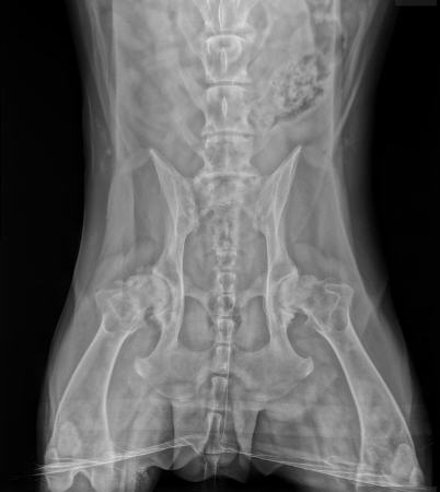
In the young dog with hip dysplasia, the earliest abnormality visible on ventrodorsal extended hip view is subluxation of the femoral head. Subluxation is defined as less than 50% coverage of the femoral head by the dorsal acetabular rim. With persistent instability and joint capsule stretching, a mineralized insertion of the joint capsule may become apparent lateral to the femoral head. This is referred to as the "Morgan's line". As laxity progresses to degenerative joint disease, remodeling of the joint is apparent. Osteophytosis, flattening of the femoral head, and shallowing of the acetabulum may be evident.

Hip Dysplasia in Dogs | Causes, Treatment & More

Radiographic sign
-Incongruency, shallow acetabulum, deformed femoral head, femoral head luxation/ subluxation, DJD, osteophytes, thickened femoral head, MORGAN LINES

https://www.dvm360.com/view/diagnosing-hip-dysplasia-proceedings
https://www.cliniciansbrief.com/article/canine-hip-dysplasia-part-1
https://veterinarypartner.vin.com/default.aspx?pid=19239&id=4952203

HIP DYSPLASIA HIP DYSPLASIA Reviewed by Dream Investor on March 29, 2020 Rating: 5

No comments:

Powered by Blogger.